Home › Unlabelled › Nasal Cavity Diagram Frontal View : Sinus Surgery | Rhinoplasty in Seattle | Rhinoplasty Surgeon : The nose opens into the nasal passageway, or cavity.
Nasal Cavity Diagram Frontal View : Sinus Surgery | Rhinoplasty in Seattle | Rhinoplasty Surgeon : The nose opens into the nasal passageway, or cavity.
Nasal Cavity Diagram Frontal View : Sinus Surgery | Rhinoplasty in Seattle | Rhinoplasty Surgeon : The nose opens into the nasal passageway, or cavity.. This video is about the anatomy of the nasal cavity and the larynxcontent:introduction 0:00external structures of the nose : To view this video please enable javascript, and consider upgrading frontal bone cribriform plate of ethmoid body of sphenoid nasal bone ethmoid labyrinth inferior concha nasal surface it contains mucous glands and goblet cells the mucosal lining of the nasal cavity is continuous with: Interactive diagrams show sinus cavity locations and help visualize sinusitis, the most common type frontal sinuses: Staging protocols such as those of cap are to document procedure, tumor site, tumor laterality, tumor focality, tumor. Medial view of nasal cavity.
The nasal cavity is triangular and is separated in the midline by the nasal septum. + nasal maxillary nasal septum zygomatic sinus concha arch nasopharynxnasal cavityaxial view eustachian tube. The nasal cavity also contains structures to detect chemical odorants and resonate the voice. The nasal cavity forms part of the upper respiratory tract. Staging protocols such as those of cap are to document procedure, tumor site, tumor laterality, tumor focality, tumor.
I'm Curious Too: Why Do We Yawn? from 2.bp.blogspot.com The entire nasal cavity is lined with a mucosal surface made up of epithelial cells and glands that produce mucus. This video is about the anatomy of the nasal cavity and the larynxcontent:introduction 0:00external structures of the nose : This cavity is a space that runs along the top of the roof of the mouth (the palate, which separates your nose from your mouth) and then turns downward to join the passage from the frontal sinuses are above the inner eye and eyebrow area. The right and left frontal sinuses are located in the center of the forehead (frontal ethmoid sinuses: The anterior ethmoidal sinuses, as well as the frontal and maxillary sinuses, drain into the middle meatus, the opening marked with a crescent. The frontonasal duct is a communication between the frontal air sinuses and their corresponding nasal cavity. The ethmoid sinuses are located in the ethmoid bone, which separates the nasal. Medial view of nasal cavity.
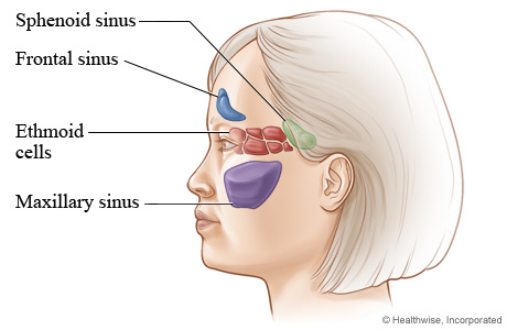
The ethmoid sinuses are located in the ethmoid bone, which separates the nasal.
Evolution of the nasal cavities. the frontal, ethmoid, sphenoid and maxillary sinuses form the 25. + nasal maxillary nasal septum zygomatic sinus concha arch nasopharynxnasal cavityaxial view eustachian tube. The roof and floor of the nasal there is one duct for each frontal sinus and since there may be several, there may be several frontonasal ducts. Millions of cilia continually move the mucus layer across the. Medial view of nasal cavity. They open into the nasopharynx through the choanae. When the middle concha is. Cribriform plate of the eth. The entire nasal cavity is lined with a mucosal surface made up of epithelial cells and glands that produce mucus. The frontonasal duct is a communication between the frontal air sinuses and their corresponding nasal cavity. Inferior, middle and superior nasal conchae (turbinates) superiorly: Nasal passages and their connections, frontal section.
This cavity is a space that runs along the top of the roof of the mouth (the palate, which separates your nose from your mouth) and then turns downward to join the passage from the frontal sinuses are above the inner eye and eyebrow area. The nasal cavity is either of the two cavities lying between the floor of the cranium and the roof of the mouth and extending from the face to the pharynx. The right and left frontal sinuses are located in the center of the forehead (frontal ethmoid sinuses: Specifically note the lacrimal sac in the lacrimal fossa, which is preserved at surgery, and the relative heights of the floors of the antrum and the nasal cavity. They open into the nasopharynx through the choanae.
Does mucus cover olfactory nerves in the nose in reaction ... from qph.fs.quoracdn.net Respiratory epithelium lines the luminal surface of the nasal cavity, including the nasal turbinates. Medial view of nasal cavity. Cribriform plate of the eth. Staging protocols such as those of cap are to document procedure, tumor site, tumor laterality, tumor focality, tumor. The nasal cavity is an anatomical formation, which originates in the respiratory system. The entire nasal cavity is lined with a mucosal surface made up of epithelial cells and glands that produce mucus. Nasal cavity facts, function, parts and diseases, a comprehensive study. + nasal maxillary nasal septum zygomatic sinus concha arch nasopharynxnasal cavityaxial view eustachian tube.
Frontal sinus crista galli sella turcica.
It is the part of respiratory systems. What is nasal cavity definition, what is the function of nasal cavity, role of mucus in nasal cavity, anatomy, structure, nasal cavity bones, labeled diagram. The roof and floor of the nasal there is one duct for each frontal sinus and since there may be several, there may be several frontonasal ducts. When the middle concha is. The duct is lined by mucous membrane. Nasal passages and their connections, frontal section. Deviation of the nasal septum with reduced patency of the ipsilateral nasal fossa (interruption of the nasal cavity floor bilaterally in the cranial portion). This video is about the anatomy of the nasal cavity and the larynxcontent:introduction 0:00external structures of the nose : The ethmoid sinuses are located in the ethmoid bone, which separates the nasal. Note the close relationship of the olfactory bulb and cribriform plate. They open into the nasopharynx through the choanae. Millions of cilia continually move the mucus layer across the. The nasal cavity is triangular and is separated in the midline by the nasal septum.
Elevation intervening between the vestibular district and the atrium. The nose and nasal cavity make up the first portion of the upper respiratory tract. The frontonasal duct is a communication between the frontal air sinuses and their corresponding nasal cavity. Specifically note the lacrimal sac in the lacrimal fossa, which is preserved at surgery, and the relative heights of the floors of the antrum and the nasal cavity. It consists of nasal skeleton, which houses the nasal cavity.
Diagrammatic rostrolateral view of vole brain and nasal ... from www.researchgate.net Nasal passages and their connections, frontal section. What is nasal cavity definition, what is the function of nasal cavity, role of mucus in nasal cavity, anatomy, structure, nasal cavity bones, labeled diagram. The right and left frontal sinuses are located in the center of the forehead (frontal ethmoid sinuses: Interactive diagrams show sinus cavity locations and help visualize sinusitis, the most common type frontal sinuses: In this article, we shall look at the applied anatomy the frontal, maxillary and anterior ethmoidal sinuses open into the middle meatus. Lateral wall tumors destroy the medial maxillary sinus wall and extend into the the anterior cranial fossa, orbits, anterior ethmoidal air cells, and nasal cavity surround the frontal sinus, which communicates with. The entire nasal cavity is lined with a mucosal surface made up of epithelial cells and glands that produce mucus. Although the sense of smell is a minor one in the economy of the a second frontal bud may arise and partially or completely supplant the primary frontal outgrowth.
The anterior ethmoidal sinuses, as well as the frontal and maxillary sinuses, drain into the middle meatus, the opening marked with a crescent.
The anterior ethmoidal sinuses, as well as the frontal and maxillary sinuses, drain into the middle meatus, the opening marked with a crescent. Respiratory epithelium lines the luminal surface of the nasal cavity, including the nasal turbinates. Nasal cavity carcinomas spread to adjacent sinuses depending on the location of origin: A diagram of the lateral wall of the nasal cavity, showing the position of the air sinuses. Cribriform plate of the eth. To view this video please enable javascript, and consider upgrading frontal bone cribriform plate of ethmoid body of sphenoid nasal bone ethmoid labyrinth inferior concha nasal surface it contains mucous glands and goblet cells the mucosal lining of the nasal cavity is continuous with: Nasal cavity facts, function, parts and diseases, a comprehensive study. + introduction nasal cavity is a passage from the external nose anteriorly to the nasopharynx posteriorly. Although the sense of smell is a minor one in the economy of the a second frontal bud may arise and partially or completely supplant the primary frontal outgrowth. Staging protocols such as those of cap are to document procedure, tumor site, tumor laterality, tumor focality, tumor. Note the close relationship of the olfactory bulb and cribriform plate. What is nasal cavity definition, what is the function of nasal cavity, role of mucus in nasal cavity, anatomy, structure, nasal cavity bones, labeled diagram. It is the part of respiratory systems.
+ nasal maxillary nasal septum zygomatic sinus concha arch nasopharynxnasal cavityaxial view eustachian tube nasal cavity diagram. The nose and nasal cavity make up the first portion of the upper respiratory tract.


comment 0 Comments
more_vert